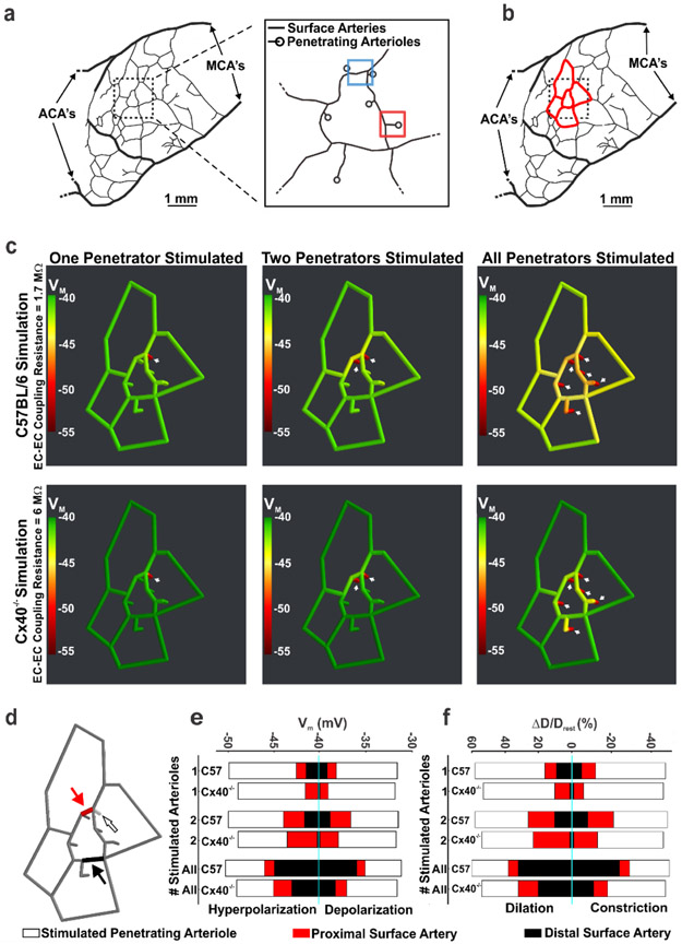
Figure 4: Vascular cells retain biophysical properties that support the ascension of responses from penetrating arterioles to surface vessels.
(a, b) Using observations from Shih et al., 2009, a network model of surface arteries and penetrating arterioles was built to study the spread of electrical/vasomotor responses. The in silico model consisted of an interconnected network of surface arteries and six adjoining penetrating arterioles. Simulations entailed voltage clamping (15 mV positive or negative to resting VM, 500 ms) one distal segment in one, two or all penetrating arterioles and resolving the voltage/vasomotor response throughout the network. (c) The ascension of hyperpolarization was color mapped along the network; endothelial-to-endothelial coupling resistance was set to 1.7 (C57BL/6 control) or 6 (Cx40−/−) MΩ (calculated in Figure 2). (d-f) One distal segment in one, two or all penetrating arterioles was/were stimulated and steady state responses (smooth muscle VM and diameter) plotted at 3 sites in the network. Those included the penetrating arteriole (200 μm upstream from the stimulation site) and two surface vessel sites. (f) D and Drest represent peak response and resting diameters, respectively. MCAs = Middle cerebral arteries; ACA’s = Anterior cerebral arteries. Figures 4 a, b are reused after permission from SAGE Publishing Ltd.

