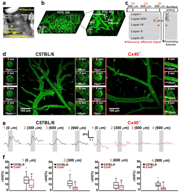
Figure 5: Compromised vascular conduction impairs neurovascular coupling responses.
(a,b) Awake mice were positioned on an exercise ball with their head fixed to an external apparatus, to facilitate two-photon imaging of the microvasculature labeled with FITC Dextran. c) Vasomotor responses to whisker stimulation were monitored at 4 sites upstream (orange numbers) near to the point of barrel cortex activation. (d) Photomicrographs of functional dilation at four vessel sites, prior to (0 s) and during (2 s) whisker stimulation in C57BL/6 and Cx40−/− mice. (e,f) Representative tracings and summary data of the functional vasomotor response to whisker stimulation. Measurements show significant attenuation of conducted responses in the penetrating arterioles and cortical surface vessels of Cx40−/− mice (0 μm: (U =85, P<0.05, Mann-Whitney U test; C57BL/6, n = 21 vessels; Cx40−/−, n = 14 vessels)), (300 μm: (U =24, P<0.05, Mann-Whitney U test; C57BL/6, n = 21 vessels; Cx40−/−, n = 14 vessels)), (600 μm: (U =126, P<0.05, Mann-Whitney U test; C57BL/6, n = 19 vessels; Cx40−/−, n = 21 vessels)), (900 μm: (U =22.50, P<0.05, Mann-Whitney U test; C57BL/6, n = 9 vessels; Cx40−/−, n = 16 vessels)). * denotes significant difference between groups.

