Fig. 2.
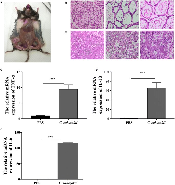
C. sakazakii induced histopathological impairment and increased pro-inflammatory cytokines levels of mammary gland in mice. a A mouse model of mastitis: the fourth pair of mammary glands were injected with C. sakazakii and the first and second pairs of mammary glands were injected with PBS and dissected. Mouse was dissected 24 h after infection. b HE staining of mammary glands histology: each nipple was injected with 100-μL C. sakazakii fluid. c HE staining of mammary glands histology: each nipple was injected with 100-μL PBS. The mRNA level of TNF-α (d), IL-1β (e), IL-6 (f), and relative mRNA level of β-actin were determined by qRT-PCR. Values are presented as means ± the standard deviation (SD) (n ≥ 3) (***P<0.001)

