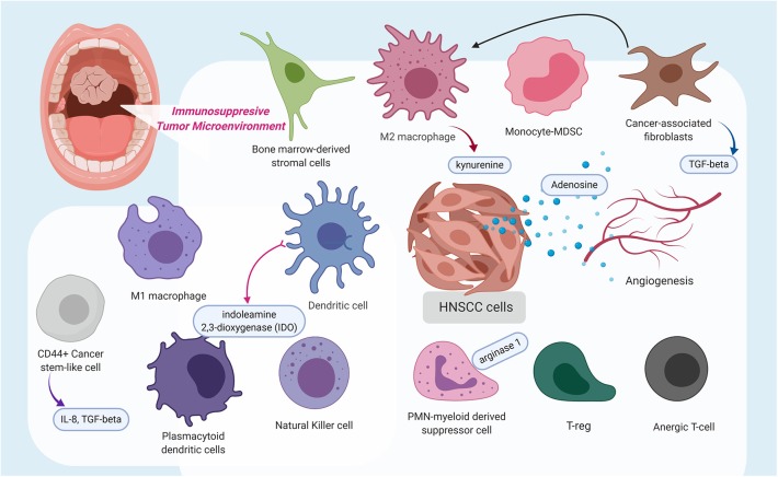
Figure 1.
This schematic diagram highlights the immunosuppressive tumor microenvironment (TME) in which a variety of immune cells are polarized to possess pro-tumoral features, stimulated cancer-associated fibroblasts, which release transforming growth factor-β, and even angiogenesis contribute to the immunosuppressive state. Additionally, several molecules, such as kynurenine, adenosine, indoleamine 2,3-dioxygenase, arginase 1, interleukin (IL)-8, and IL-10, were identified to contribute to a pro-tumoral immunosuppressive TME or at extreme, an immune-desert. The figure was created with BioRender.com and was exported under a paid subscription.

