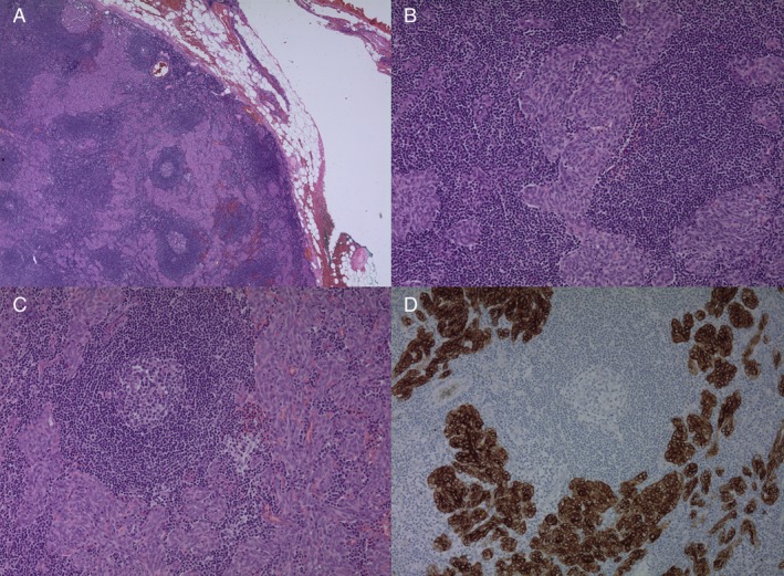
Figure 2.

Low‐magnification view shows multiple small nodules of tumour cells separated by abundant lymphoid stroma (A). The tumour cells are bland‐looking, short spindled and oval, epithelial cells (B). The intervening lymphoid stroma contains lymphoid follicles with germinal centres (C). Immunohistochemistry for pancytokeratin is positive in tumour cells, confirming the diagnosis of thymoma (D).
