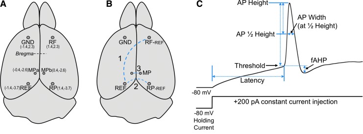
FIG. 1.
(A) Dorsal view of mouse brain showing positions of electrode placement. Parentheses indicate absolute position in millimeters relative to medial and bregma (X,Y). The ground electrode (GND) was placed in the left frontal lobe. A reference electrode (REF, in left parietal region) served to record from right frontal (RF) and right parietal (RP) regions. Two electrodes in the medial parietal (MPa and MPb) region served as a bipolar electrode. (B) Recording electrode pairs are indicated by a dashed blue line. (C) Diagram of action potential parameters measured from a 200 pA constant current injection added to a −80 mV holding current. A description of these parameters are detailed in Methods.

