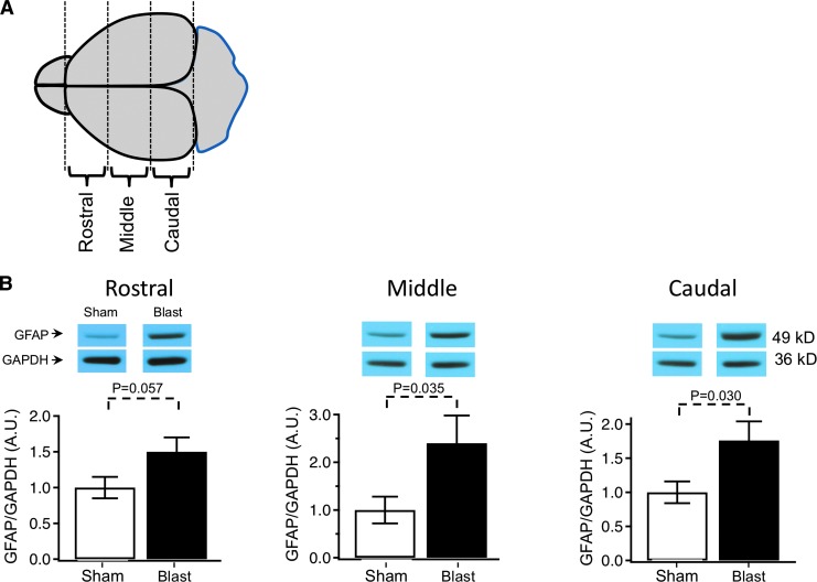
FIG. 2.
(A) Diagram indicating regions of the brain analyzed for Western blot detection. The brain sections included 3 mm rostral, middle, and caudal portions. (B) Glial fibrillary acidic protein (GFAP) expression after repetitive (3X) blast injury measured in the rostral, middle, and caudal regions of the brain. Expression is normalized to glyceraldehyde 3-phosphate dehydrogenase (GAPDH) expression, and to sham control: rostral: 1 ± 0.15 sham, 1.50 ± 0.20 traumatic brain injury (TBI); middle: 1 ± 0.28 sham, 2.4 ± 0.58 TBI; caudal: 1 ± 0.16 sham, 1.76 ± 0.28 TBI, n = 12 animals for sham and 12 blast TBI. The p values are indicated in bar graphs.

