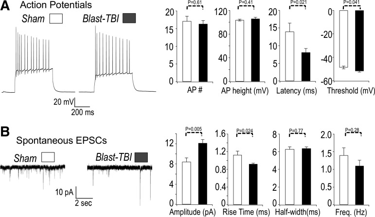
FIG. 6.
(A) Left panel. Sample action potential (AP) recordings from sham-treated (sham) mouse and mice that received three repetitive blast injuries (blast). The APs were measured using whole cell recordings of dentate gyrus granule neurons. The APs were evoked by a 200 pA current injection. Right panel, summary of data. The AP number (per 500 msec current injection): 17.1 ± 1.3 sham, 16.3 ± 0.9 traumatic brain injury (TBI); AP height: 103.7 ± 1.5 mV sham, 106.0 ± 2.4 mV TBI; AP latency (time between current injection and first spike): 14.0 ± 2.6 msec sham, 8.1 ± 1.1 msec TBI; AP threshold: −48.6 ± 1.2 mV sham, −51.7 ± 0.9 mV TBI. n = 25 sham, 32 blast TBI. (B) Blast TBI increases the amplitude and decreases rise time of EPSCs. Left panel. Sample recording of spontaneous excitatory post-synaptic currents in dentate gyrus granule neurons. The excitatory post-synaptic currents (EPSCs) were measured at −80 mV holding potential. Right panel, summary of data. EPSC amplitude: 8.4 ± 0.8 pA sham, 12.1 ± 0.7 pA TBI; EPSC rise time: 1.1 ± 0.08 msec sham, 0.92 ± 0.02 msec TBI; half-width: 6.3 ± 0.3 msec sham, 6.4 ± 0.2 msec TBI. n = 177 events sham, 227 events TBI for above EPSC data. Frequency 1.4 ± 0.2 Hz sham, 1.1 ± 0.1 Hz TBI, n = 10 sham, 15 TBI for frequency data. p values are indicated on the bar plots.

