Figure 10.
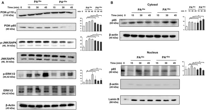
Cellular signaling pathways were altered in J774A.1 macrophages in response to PA−Sia/PA+Sia infection. (A) J774A.1 were infected with PA−Sia or PA+Sia for 15, 30, or 45 min. A few molecules involved in cellular signaling pathways were detected by Western blotting. After visualization of phosphorylated forms of JNK, ERK, the same blots were stripped and processed to detect its total counterparts. (B) The expression levels of NF-κB pathway molecules and nuclear translocation of p65 subunit was assessed in cytosolic and nuclear fractions of infected macrophages. Western blots shown are representative of at least three independent experiments. Densitometric values reported are normalized with respect to β-actin band intensities. Densitometric values from at least three independent experiments were used to calculate average values of band intensity which are presented as bar diagrams with statistical significance.

