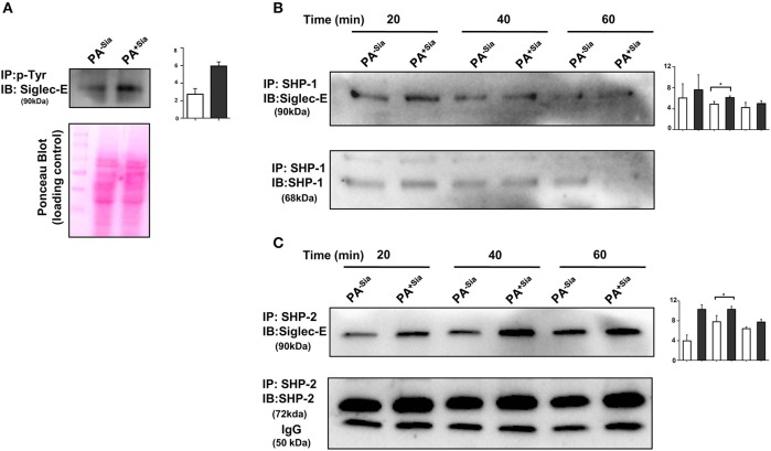
Figure 7.
Interaction of siglec-E-sialic acids results in siglec-based inhibitory signaling. (A) Phosphorylation of siglec-E was checked by immunoprecipitation of lysates of PA−Sia/PA+Sia infected macrophages with biotinylated anti-phosphotyrosine antibodies using avidin-agarose beads. Precipitated proteins were resolved via SDS-PAGE, transferred to PVDF membrane and probed for siglec-E using antibodies. Image of Ponceau S stained membrane has been used to account for equal loading of proteins. (B,C) Association between SHP-1 and SHP-2 with siglec-E was quantified by immunoprecipitating infected macrophage lysates with anti-SHP-1 or SHP-2 antibody and protein A- sepharose 4B beads. Precipitated complexes were analyzed by western blotting. All western blots are representative of three independent experiments. Densitometric values reported are normalized with respect to SHP-1/SHP-2 band intensities. Average of normalized band intensities from three independent experiments are represented as bar diagrams with statistical significance.

