
Figure 4.
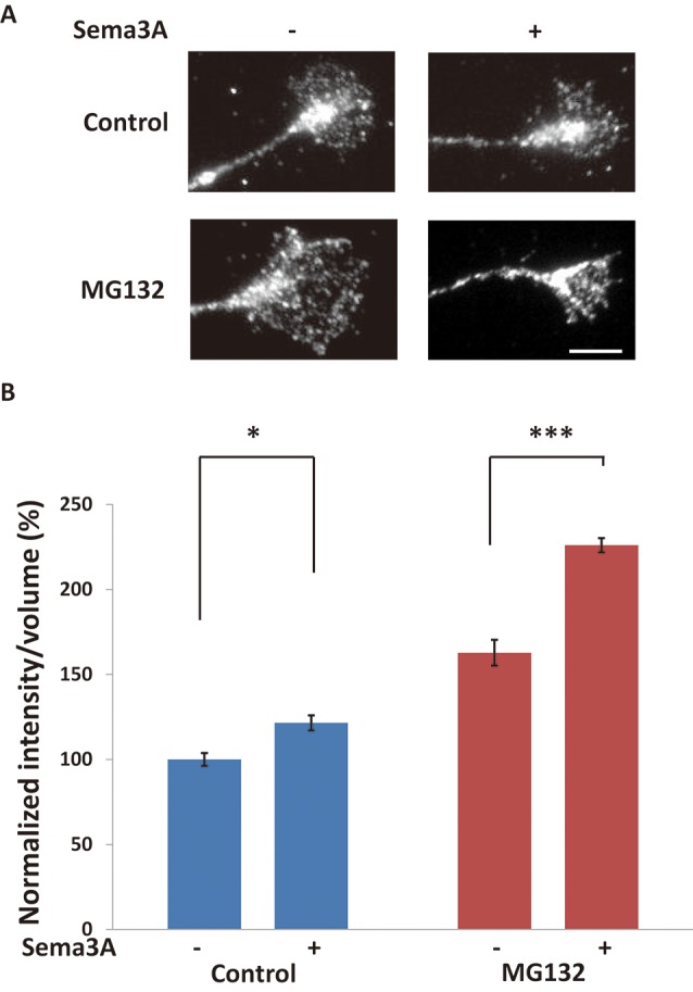
Sema3A induced protein ubiquitination in growth cones. (A) Immunofluorescence images of anti-ubiquitinated protein antibody (FK2) staining in growth cones. Sema3A treatment for 5 min increased the fluorescence intensity of ubiquitinated proteins in growth cones. MG132 enhanced the intensities of ubiquitinated proteins in growth cones stimulated by Sema3A more significantly than controls. Scale bar: 10 μm. (B) Quantification of fluorescence intensities of ubiquitinated proteins in growth cones as in (A). Data represent mean ± SEM for n = 10. *p < 0.05, ***p < 0.001, significantly different from Sema3A (-) using two-way ANOVA with Tukey’s multiple comparison test.