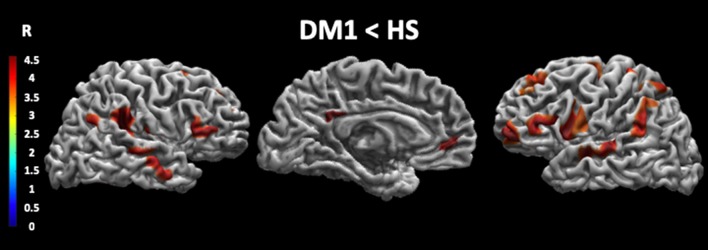
Figure 1.
Cortical thickness in DM1 patients compared with HS. This figure reveals that patients with DM1 compared to healthy subjects showed a significant decrease in cortical thickness in several brain areas. In particular, the cortical thickness is significantly decreased in the precuneus, the angular gyrus, the superior temporal gyrus and the medial frontal gyrus bilaterally, the right precentral gyrus, the right posterior and anterior cingulate cortex, and the left superior parietal lobule. The statistical comparisons were overlapped on MRIcron ch2bet template (https://www.nitrc.org/projects/mricron). DM1, myotonic dystrophy type 1; HS, healthy subjects; R, right.

