
Figure 3.
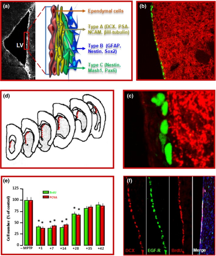
Location, proliferation and dopaminergic innervation of SVZ‐NSCs. (a) Microscopic brain image at the level of the striatal SVZ. In the inset, a schematic drawing of the four SVZ‐cell types: 1. slowly dividing SVZ astrocytes (type B cells), 2. rapidly dividing transit‐amplifying cells (type C cells, TAPs), 3. migrating neuroblasts (type A cells), and 4. ependymal cells (type E cells) (Doetsch et al., 1997, 1999). (b, c) Nigrostriatal DAergic neurons originating in the SN project to the SVZ. Dual immunofluorescent staining with dopamine transporter (DAT, in red) and bromodeoxiuridine (BrdU, green) showing a dense network of DAT expressing neurons innervating the SVZ (b; and higher magnification in c). (d–f) schematic representation of cell counting performed in coronal sections through the SVZ (d); stereological estimations of BrdU and proliferating cell nuclear antigen (PCNA)‐positive cells (e), and representative stainings of DCX+, EGF‐R+, BrdU+, counterstained with the nuclear marker 4,6‐diamidino‐2‐phenylindole, Dapi (blue) (f), indicating that MPTP‐induced basal ganglia injury resulted in a biphasic time‐dependent response: a down‐regulation of SVZ‐NSC proliferation followed by a return to pre‐MPTP levels