
Figure 2.
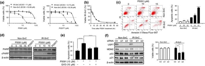
Inhibition of USP7 activity selectively induces apoptosis in SnCs. (a) Effect of USP7 inhibitors on cell viability of non‐SnC and IR‐SnC WI‐38 cells. Viability was determined after cells were treated with the indicated concentrations of P5091 or P22077 for 72 hr. Data are presented as mean ± SEM (n = 3). (b) P5091 induces cell death in IR‐SnC in a time‐dependent manner. IR‐SnC WI‐38 cells were treated with 15 µM P5091, and cell viability was determined at indicated time points. Data are presented as mean ± SEM (n = 4). (c) P5091 selectively induces apoptosis in IR‐SnC in a dose‐dependent manner. Apoptosis was determined after cells were treated with P5091 for 72 hr by Annexin V‐Alexa Fluor 647 and PI staining and flow cytometry. Representative flow cytometric plots are shown in the left panel, and percentages of apoptotic cells (Annexin V+ and Annexin V+/PI+ cells) are presented in the right panel as mean ± SEM of 3 independent experiments. *p < .05 and ****p < .0001. (d) Representative images of Western blots of PARP and cleaved PARP (cPARP) in non‐SnC and IR‐SnC WI‐38 cells 72 hr after treatment with P5091. (e) Percentages of apoptotic cells in IR‐SnC WI‐38 cells after pretreatment with the pan‐caspase inhibitor QVD for 4 hr, and then with or without P5091 for 24 hr. Data are presented as mean ± SEM (n = 3). ****p < .0001. (f) Knockdown of USP7 expression by siRNA reduces MDM2, upregulates p53, and induces apoptosis in IR‐SnCs. USP7, p53, and MDM2 protein levels were determined after transfection with control siRNA (Ctr), USP7 siRNA‐1 (si1), or USP7 siRNA‐2 (si2) for 48 hr. Cell viability in non‐SnC and IR‐SnC WI‐38 cells was determined 4 d after transfection. Representative images of Western blots are shown in the left panel, and percentages of viable cells are presented in the right panel as mean ± SEM of 3 independent experiments. **p < .01