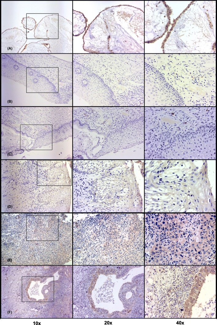
Fig. 1.
Representative immunohistochemical staining pattern of B7-H6 in cervical precancerous lesions and invasive cervical cancer. Specimens were categorized as follows: (a) ovarian cystadenoma (as the positive control): B7-H6 is mainly expressed in lining epithelium; (b) cervicitis: absence of B7-H6 in keratinocytes; (c) LSIL: absence of B7-H6 in abnormal keratinocytes; (d) HSIL: presence of B7-H6 in abnormal keratinocytes; (e) SCC: B7-H6 expression in transformed tumoral cells; (f) UCAC: strong expression of B7-H6 in transformed tumoral cells. Positive staining of B7-H6 is seen in the cytoplasm and membrane of transformed cells, and in plasma cells and mononuclear cells infiltrating the stroma. Photomicrographs were taken with different objectives (10x, 20x, and 40x). Abbreviations: LSIL: low-grade squamous intraepithelial lesion; HSIL: high-grade squamous intraepithelial lesion; SCC: squamous cervical carcinoma; UCAC: uterine cervical adenocarcinoma

