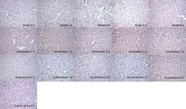
Figure 2.
Effect of acupuncture on brain histomorphology in rats with cerebral hemorrhage, assessed by hematoxylin-eosin staining.
In the control group, brain tissue was intact, and neurons and glial cells appeared normal (arrow). In the model group, brain tissue showed hemorrhagic foci at 6 hours and edema at 1 and 2 days, and vacuoles of different sizes were visible (arrows). Numbers of dema and vacuoles were the greatest at 3 days (arrows). The nuclei of the neurons around the focus were condensed and stained (arrows), and the edema around the 7-day hematoma tissue was less (arrows). In the acupuncture group, the brain tissue showed hemorrhagic foci at 1 day, but the cellular morphology appeared more normal (arrows). At 3 days, brain tissue around the hematoma appeared improved, the degree of edema was less (arrows), and the number of inflammatory cells was reduced (arrows). Brain edema had resolved at 7 days. Original magnification: 400×.

