Figure 1.
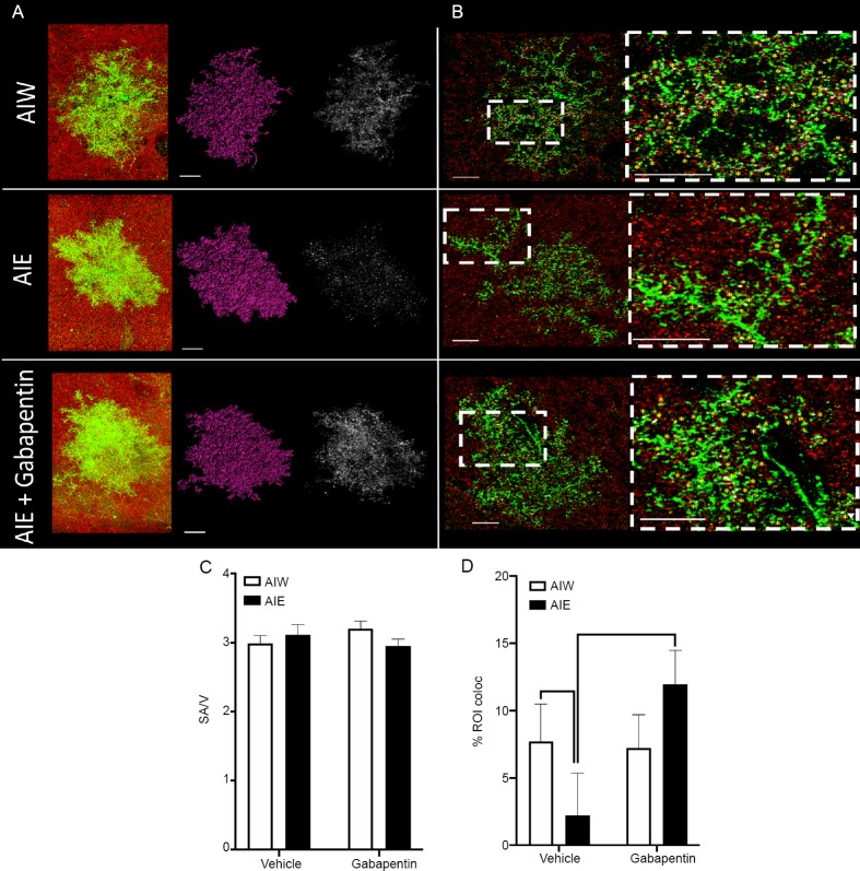
AIE-induced reduction of astrocyte-synapse proximity in hippocampal area CA1 is reversed by gabapentin.
(A) Left to right: Raw z-series astrocytes (green) and synaptic puncta (red), rendered astrocytes (purple), and points of colocalization of the astrocyte and synaptic maker (white), from control animals that received AIW, those exposed to AIE, and AIE animals that received gabapentin in adulthood (AIE + gabapentin). Scale bars: 15 μm. (B) Left Column: Astrocytes (green) and synaptic puncta (red) from each of the treatment groups described under A. Inset boxes represent regions for expanded magnification. Scale bars: 15 μm. Right column: Expanded magnification of areas inset in left column. Yellow asterisks indicate identified points of astrocyte-synaptic colocalization. Scale bars: 15 μm. (C) Mean (± SEM) ratio of astrocyte surface area (SA) to volume (V) was unchanged by AIE or Gabapentin treatment. (D) Mean (± SEM) percent of colocalization of astrocyte viral labeling and synaptic puncta was reduced in hippocampal area CA1 in adulthood following AIE (P < 0.05, vs. AIW), and this decrease was reversed by treatment with gabapentin (P < 0.05, vs. AIE and P > 0.05, vs. AIW + gabapentin). The number of animals used for each treatment group was: AIW – n = 6, AIE – n = 6, AIW + gabapentin – n = 8, AIE + gabapentin – n = 7. An average of five cells per animal were included in the mixed linear modeling analysis. AIE: Adolescent intermittent ethanol; AIW: adolescent intermittent water.

