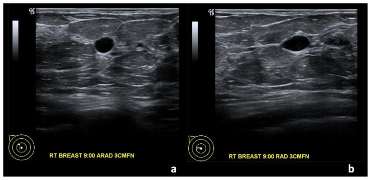
Figure 1.
67-year-old female with right breast invasive ductal carcinoma and ductal carcinoma in-situ. Two years prior to Figures 2 and 3.
Findings: (a, b) The right breast demonstrates an oval, circumscribed, parallel, anechoic mass with posterior acoustic enhancement at the 9 o’clock position, 3 cm from the nipple, measuring 0.8 × 0.5 × 0.4 cm, consistent with a simple cyst.
Technique: Outside hospital ultrasound examination using a high frequency linear transducer (14 MHz).

