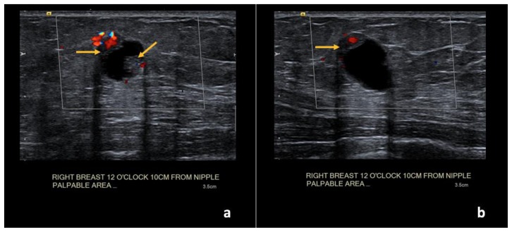
Figure 11.
73-year-old female with right breast infiltrating duct carcinoma.
Findings: (a, b) The right breast demonstrates an oval, circumscribed, complex solid and cystic mass at the 12 o’clock position, 10 cm from the nipple, measuring 1.6 × 1.1 × 1.2 cm with new vascular internal mural nodularity (arrows).
Technique: Ultrasound and Color Doppler examination using a high frequency linear transducer (12 MHz).

