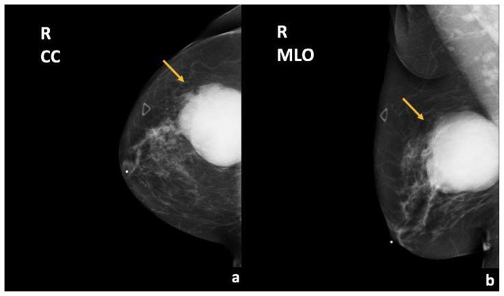
Figure 2.
67-year-old female with right breast invasive ductal carcinoma and ductal carcinoma in-situ.
Findings: There is a partially imaged oval mass with obscured margins in the upper outer quadrant (arrows), corresponding to the patient’s right breast enlarging palpable abnormality.
Technique: Diagnostic mammogram (30 kVp, 78 mAs). (a) Craniocaudal (CC) and (b) mediolateral oblique (MLO) views of the right breast.

