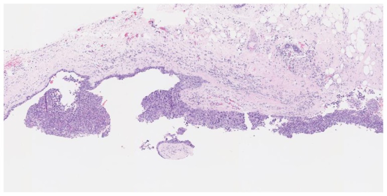
Figure 4.
67-year-old female with right breast invasive ductal carcinoma and ductal carcinoma in-situ.
Findings: The cystic lesion demonstrates infiltrating poorly differentiated duct carcinoma, grade 3 of 3, Nottingham score of 8. The tumoral cells exhibit increased mitosis, nuclear pleomorphism and no tubule formation, indicating high grade malignancy.
Technique: Microscopic examination with Hematoxylin and Eosin (H&E) stain. Medium-power view (55×) from the core needle biopsy of a right breast complex solid and cystic mass.

