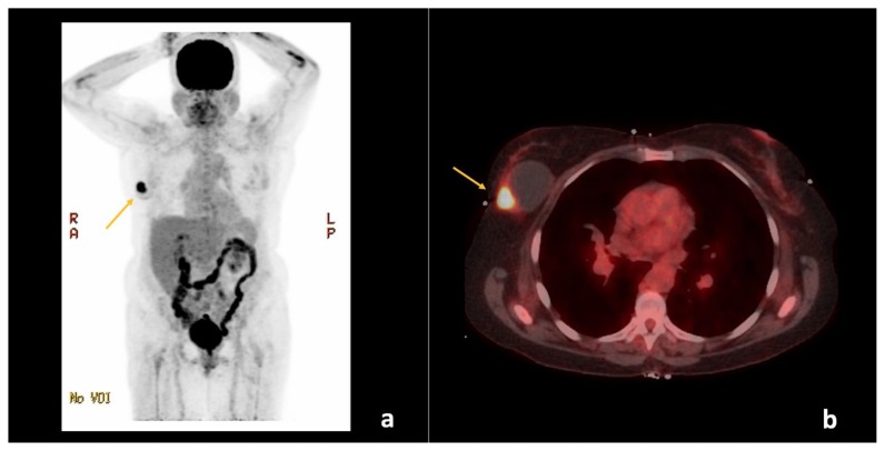
Figure 5.
67-year-old female with right breast invasive ductal carcinoma and ductal carcinoma in-situ.
Findings: There is a hypermetabolic focus within the right breast with maximum standardized uptake value (SUVmax) of 13.7 (arrows), corresponding to the patient’s known primary malignancy. No evidence of regional or distant metastases.
Technique: Positron emission tomography-computed tomography (PET/CT) from the skull to the mid-thigh level. (a) Maximum intensity projection (MIP) and (b) Fused axial plane images through the upper chest approximately 67 minutes following intravenous injection of 12.268 mCi of 18F-2-fluoro-2-deoxyglucose (F18-FDG) through the patient’s right antecubital vein. The patient’s fasting blood glucose measured just before intravenous injection was 75 mg/dL. The low-dose CT was with oral contrast, but without intravenous contrast and was used for image fusion, attenuation correction, and lesion localization only. The standardized uptake values (SUVs) are normalized to the patient’s body weight and indicate the highest activity concentration (SUVmax) in a given disease site. (GE Medical Systems, Discovery 690; 140 kVp, 18 mA, 3.75 mm slice thickness. DFOV of MIP: 65.0 × 130.0 cm. DFOV of Fused Axial image: 50.0 cm).

