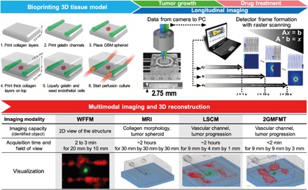
Fig. 1. Conceptual figure for the workflow.

(Top) Integration of the platform: 3D tumor tissue model and 2GMFMT. The platform enables long-term tissue culture and longitudinal assessment of tumor invasion. Bio-printing: 3D tissue containing a GBM spheroid and vascular channels were bioprinted within a plexiglass perfusion chamber and cultured with medium perfusion (with or without drug). Longitudinal imaging is conducted through a thick plexiglass for both tumor growth period and drug regimen. Noninvasive imaging was conducted through a transparent plexiglass chamber. Descanned configuration enabled dense sampling of the target. Each scan point (i.e., p, r, and s) represents a pixel in the raw data. As a representative, one detector raw datum was shown. A full frame was completed when raster scanning is finished (scan point number, s). Typically, a full field of view scanning is completed in ~20 s. (Bottom) Multimodal imaging and 3D reconstruction for all potential modalities. 2GMFMT imaging was performed on the model every 3 to 4 days with 2GMFMT. 2GMFMT presents superior data acquisition time for volumetric assessment of GBM brain tumor in comparison to its counterparts microscopic magnetic resonance imaging (μMRI) and laser scanning confocal microscopy (LSCM). WFFM, wide-field fluorescence microscopy. Photo credit: Vivian Lee, Northeastern University.
