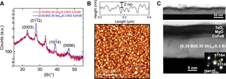
Fig. 1. Structural characterization of Bi|Sb multilayers.

(A) X-ray diffraction (XRD) spectra of 0.5 Ta/[0.35 Bi|0.35 Sb]N/0.3 Bi/2 CoFeB/2 MgO/1 Ta with N = 8 (blue line) and N = 16 (red line). a.u., arbitrary units. (B) AFM image of the N = 8 film. A line profile along the blue solid line drawn in the bottom image is shown at the top. (C) Cross-sectional HAADF-STEM images of the N = 16 structure. Selected nanobeam diffraction pattern of the Bi|Sb multilayer is shown in the inset.
