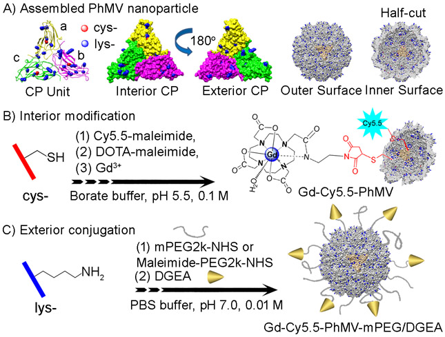
Figure 1.
(A) Structure of the Physalis mottle virus (PhMV) coat protein (CP) consisting of a (yellow), b (magenta), and c (green) subunits; the protein subunits are identical; a–c denotes their position within the icosahedral T = 3 lattice. The surface-exposed residues highlighted as external lysine (blue) and internal cysteine residues (red). Representation of interior and exterior (with 180° rotation) of the asymmetric PhMV CP subunit. The structure of the assembled hollow capsid shown as outer and inner surfaces as whole sphere and half-cut view, respectively. Methodology for (B) internal and (C) external modifications, respectively. Images were created using UCSF Chimera software (PDB: 1E57) and ChemDraw v15.0.

