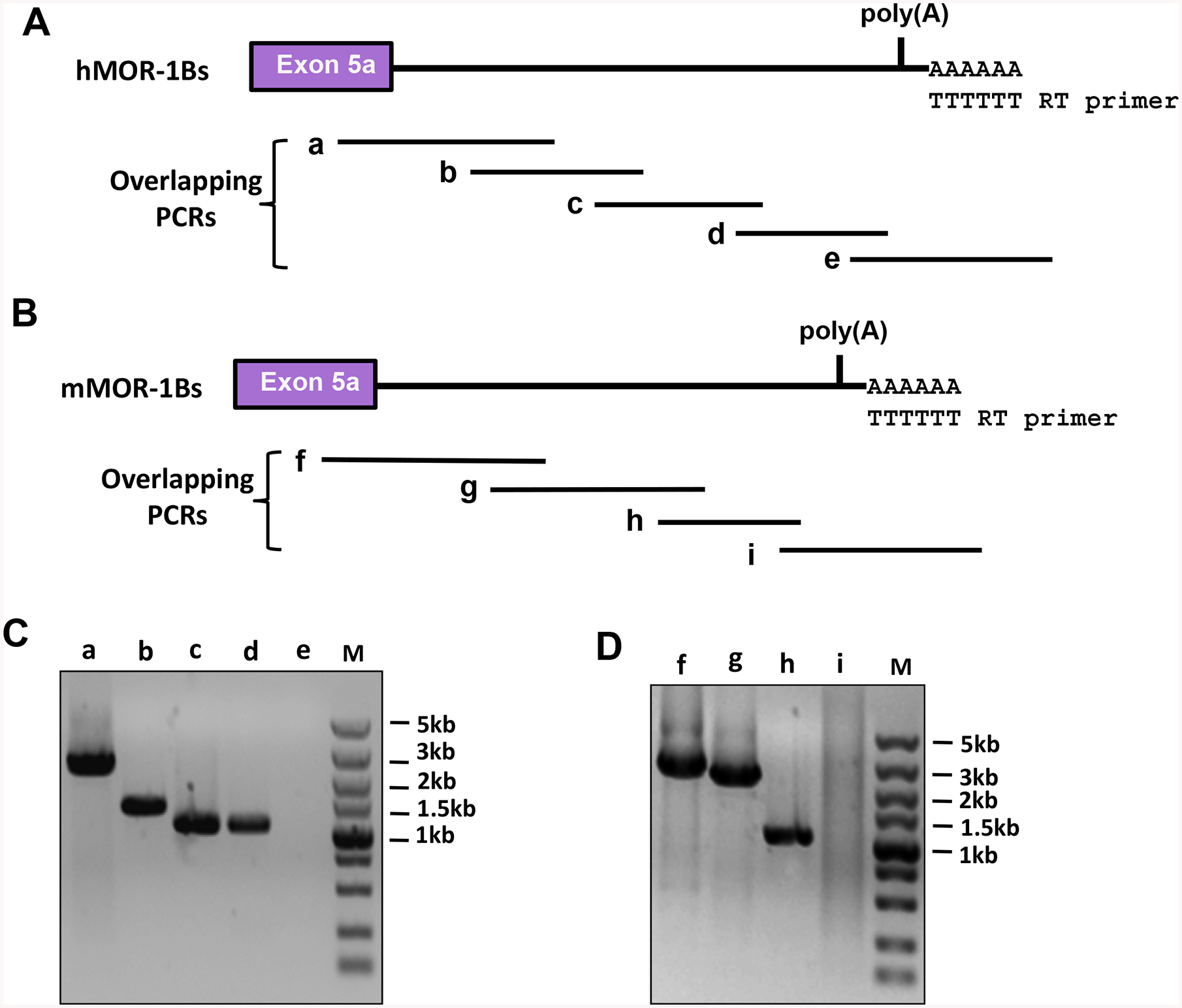
Fig. 3.

Identification of the 3’-UTR of the mouse and human MOR-1Bs using overlapping PCRs. A). Human MOR-1Bs 3’-UTR. Overlapping PCRs were performed using five pairs of primers and the first-stand cDNAs reverse-transcribed from poly(A) RNAs of Be(2)C cells to amplify overlapped PCR fragments covering the 3’-UTR, as described in Materials and Methods. a, b, c, d and e are the predicted overlapping PCR fragments.
B). Mouse MOR-1Bs 3’-UTR. Overlapping PCRs were performed using four pairs of primers and the first-stand cDNAs reverse-transcribed from total RNAs of mouse brain to amplify overlapped PCR fragments covering the 3’-UTR, as described in Materials and Methods. f, g. h and I are the predicted overlapping PCR fragments.
C & D). Analysis of the PCR products. The PCR products from Be(2)C (C) and mouse brain (D) were separated on 1% agarose gels that were stained with ethidium bromide and imaged with ChemiDoc MP System. The sizes of a – d and f – h PCR fragments were consistent with the predicted sizes (See Materials and Methods) and confirmed by DNA sequencing. PCR e and i did not yield any PCR bands.
