
Extended Data Figure 5: ILC2s induce antigen-specific CD8+ T cell priming.
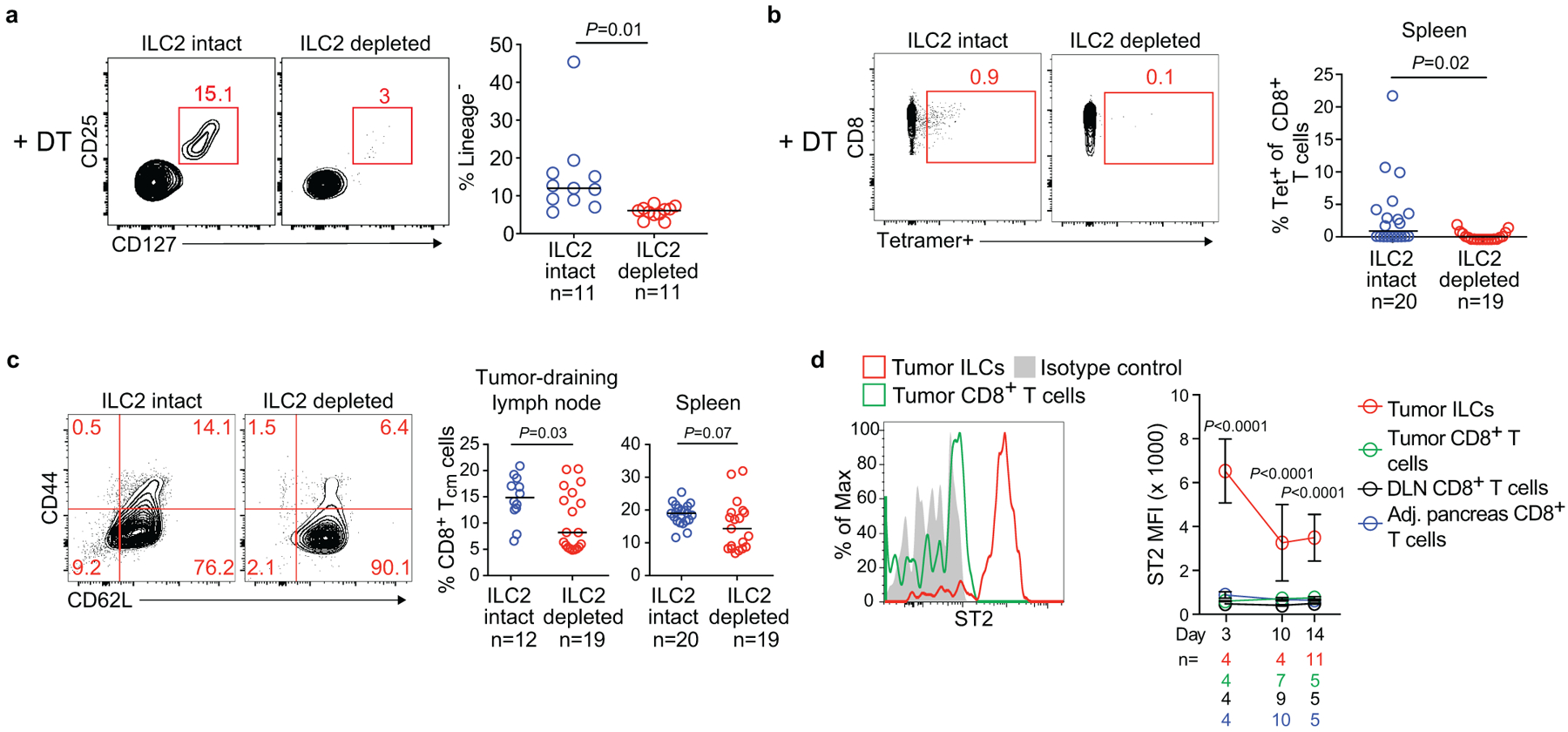
(a) Gating and frequency of intratumoral ILC2s in ILC2-intact mice (diphtheria toxin [DT]-treated Icos+/+; CD4Cre/+) and ILC2-depleted mice (DT-treated Icosfl.DTR/+; CD4Cre/+). (b) Gating and frequency of OVA-specific CD8+ T cells in spleens from ILC-intact and ILC-depleted mice. OVA-specific T cells were detected as SIINFEKL-tetramer+ cells. (c) Gating and frequency of central memory CD8+ T (TCM) cells (CD45+CD3+CD8+ CD44+CD62L+) in tumor draining lymph nodes and spleens in ILC-intact and ILC-depleted mice. (d) ST2 expression on CD45+CD3+CD8+ T cells after tumor implantation in PDAC mice. Data were collected at 14 days post tumor implantation or at the time points indicated. DLN, draining lymph node; MFI, mean fluorescence intensity. Horizontal bars mark medians; error bars mark s.e.m. n indicates individual mice analyzed separately in at least two independent experiments with n≥2/group. P values determined by two-tailed Mann-Whitney test (a-c) and two-way ANOVA with Tukey’s multiple comparison post-test (d, indicating comparison of tumor ILCs to all other groups).