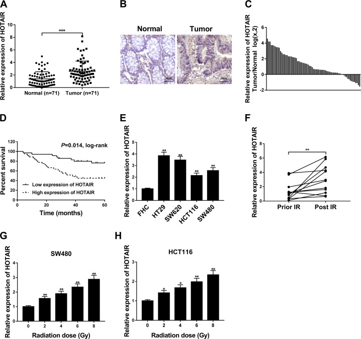
Fig. 1. HOTAIR was highly expressed in CRC tissues and cells, and HOTAIR expression was markedly upregulated after IR treatment in CRC.
a, c HOTAIR level was determined by RT-qPCR assay in 71 pairs of CRC tissues and corresponding normal tissues. b Representative images of ISH analysis for HOTAIR in CRC tissues and adjacent normal tissues. d Kaplan–Meier survival analysis of 71 CRC patients according to the difference of HOTAIR expression. e HOTAIR expression was measured by RT-qPCR assay in FHC, HT29, SW620, HCT116, and SW480 cells. f HOTAIR level was measured by RT-qPCR assay in plasma samples of 12 CRC patients before and after radiotherapy. g, h HOTAIR level was detected in SW480 and HCT116 cells treated with different doses (0, 2, 4, 6, or 8 Gy) of IR. *P < 0.05.

