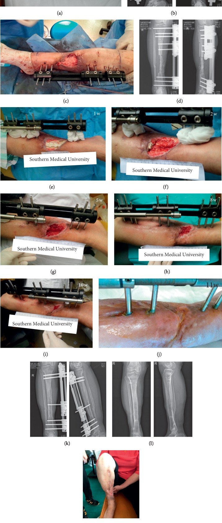
Figure 3.
(a) The appearance of the affected limb at transfer to our department; (b) X-ray of tibia and fibula; (c) after debridement and replacement of the external fixator, the bone defect site was filled with a calcium sulfate spacer loaded with vancomycin, exposing the wound surface; (d) postoperative X-ray; (e–j) the appearance of the affected limb after operation. The wound surface gradually decreased and healed throughout the bone transport process; (k) postoperative X-ray at 11 months showed that the fracture was well healed and the external fixator was removed; (l-m) both the X-ray and the appearance at 24 months after the operation.

