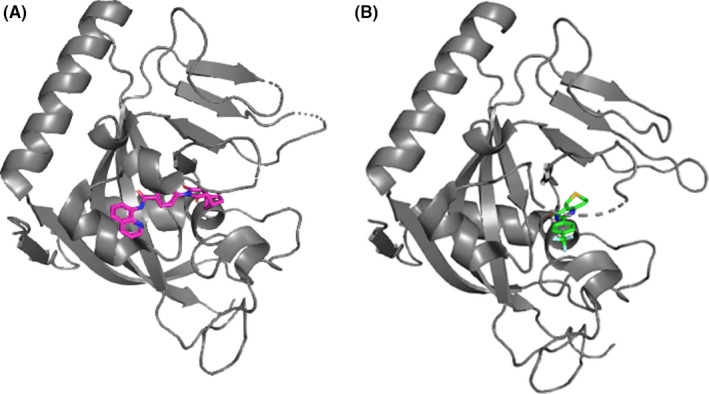
Figure 3.

Crystal structures of tankyrase‐1 (TNKS1) in complex with 2 small molecule compounds, IWR1 (A) and XAV939 (B). Data taken from RCSB Protein Data Bank (IWR1, PDB ID:4OA7 and XAV939, PDB ID:3UH4) and visualized using PyMOL molecular graphics software (https://pymol.org/2/). TNKS1, IWR1, and XAV939 are colored by gray, magenta, and green, respectively
