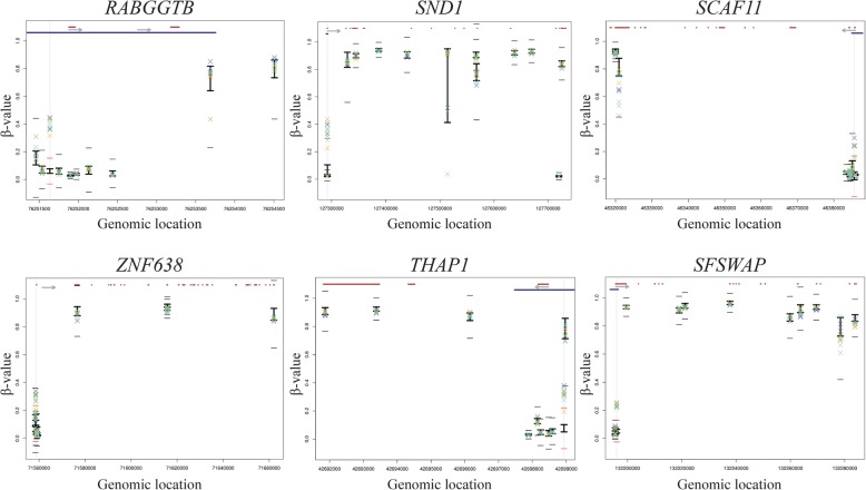
Fig. 2.
Methylation levels (β value) in all 6 significant upregulated CpGs located in the promoter regions of the genes. In the upper part of each plot, the gene structure is represented in red and promoter region (“Promoter_associated” feature retrieved from Illumina annotation) in blue. The light grey arrow represents the transcription direction of the gene. For each CpG, the BIOS values are represented by the black vertical line with upper (average + 1 SD) and lower limits (average − 1SD). The families are represented as a X of different colours (family I—green, family II—blue, family III—yellow, family IV—light purple, family V—dark blue). To be considered as significantly different from the BIOS, the families’ symbols must go beyond the small black horizontal line (average ± 5.65 SD). Genes with more than 10 CpG sites assessed by 450K array were represented by 10 randomly selected CpGs. The upregulated CpG in each plot is aligned with a vertical light grey line, and in this case, the little horizontal lines become red since the families’ symbols exceeded these limits. An extended version of figure 2 is available as supplementary information

