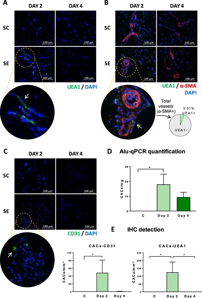
Fig. 2.
Detection of human cells in CAC-treated mice by IHC and qPCR. Human cells were analyzed by IHC in the frontal muscles of SE left limbs on days 2 and 4: a tissues infused with UEA1 pre-labeled cells, or tissues incubated with b UEA1 and anti-α-SMA antibody and c anti-hCD31, to determine the location of CACs in the tissue. d Graphical representation of the number of human cells (number of cells/μm2) detected after IHC labeling with anti-hCD31 and UEA1. e Quantification of human cells (number of cells/milligram of the tissue) by qPCR using human-specific Alu sequences, with the SH group as the negative control. Data represented as mean ± SEM, n:4 per day/group. Significant differences were seen by Kruskal-Wallis test and Dun’s test for multiple comparisons. *p value < 0.05

