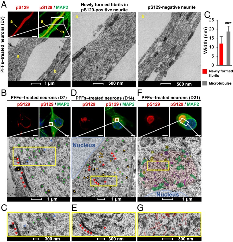
Fig. 2.
The formation and maturation of LB-like inclusions require the lateral association and the fragmentation of the newly formed α-syn fibrils over time. At the indicated time, PFF-treated neurons were fixed, ICC against pS129-α-syn was performed and imaged by confocal microscopy (A, B, D, and F, Top), and the selected neurons were then examined by EM (A, B, D, and F, Bottom). (A, a) Neurite with pS129-positive newly formed α-syn fibrils. (A, b) A neurite negative for pS129 staining. (A, c) Graph representing the mean ± SD of the width of the microtubules compared to the newly formed fibrils at D7. **P < 0.001 (Student’s t test for unpaired data with equal variance), indicating that this parameter can be used to discriminate the newly formed fibrils from the cytoskeletal proteins. (Scale bars: A, B, D, and F, Top, 10 μm.) (C–G) Representative images at higher magnifications corresponding to the area indicated by a yellow rectangle in EM images shown in B, D, and F.

