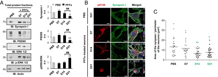
Fig. 7.
Synaptic dysfunctions were associated with the formation and maturation of the LB-like inclusions. (A) The levels of Synapsin I, PSD95, and ERK 1/2 were assessed by Western blot over time. Actin was used as the loading control. The graphs represent the mean ± SD of a minimum of three independent experiments. (B and C) Synaptic area decreases in PFF-treated neurons from D14. (B) Aggregates were detected by ICC using pS129 (MJFR13) and Synapsin I antibodies. Neurons were counterstained with MAP2 antibody, and the nucleus with DAPI. (Scale bars: 5 μm.) (C) Measurement of the synaptic area was performed over time. (A and C) ANOVA followed by Tukey honest significant difference [HSD] post hoc test was performed. *P < 0.05, **P < 0.005 (PBS- vs. PFF-treated neurons). ##P < 0.005 (D14 vs. D21 PFF-treated neurons). a.u., arbitrary unit.

