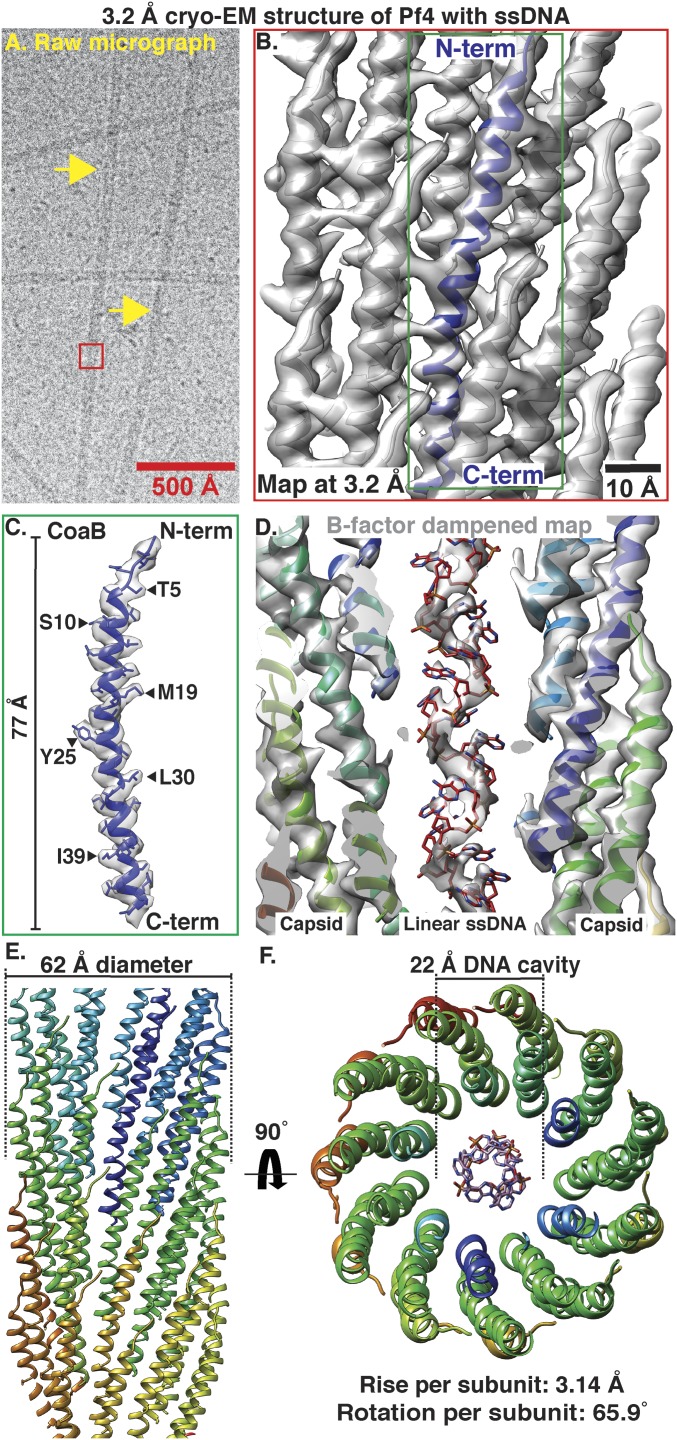
Fig. 1.
Cryo-EM structure of Pf4 phage at 3.2-Å resolution. (A) Cryo-EM image of native Pf4 phage (yellow arrows) purified from biofilms (red box represents reconstructed segment). (B) Single-particle cryo-EM reconstruction of Pf4 phage (Movie S1). Cryo-EM density is shown as a gray isosurface, with the refined atomic coordinates of Pf4 CoaB protein subunits shown as ribbons (N- and C terminus of one CoaB marked). (C) Density and atomic model for a single CoaB protein. (D) Cross-section through the cryo-EM structure shows that the Pf4 ssDNA genome is linear. Map is B-factor dampened (50 Å2 compared to B and C) to aid visualization of the ssDNA. (E) Side view of Pf4 showing interdigitated arrangement of CoaB subunits within the capsid coat (only the front CoaB subunits displayed). (F) Top view of Pf4 showing linear ssDNA within the 22-Å inner cavity.

