Fig. 3.
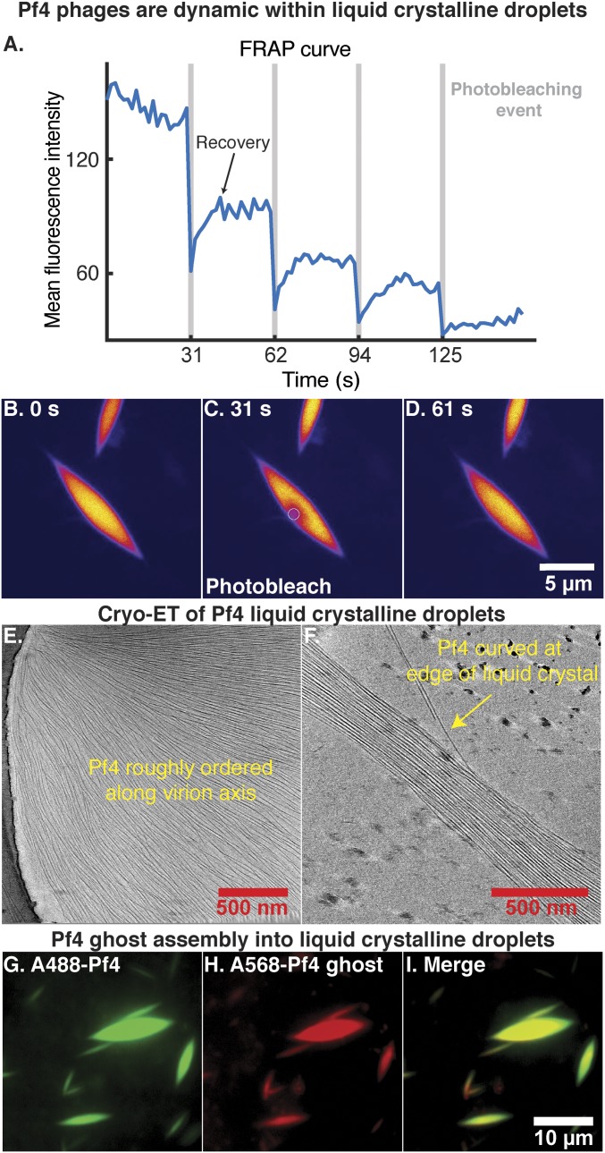
Pf4 assembly into liquid crystalline droplets is dynamic with strong orientational ordering of filaments. (A) A region within a liquid crystalline droplet was photobleached multiple times and the FRAP was measured over time (Movie S3). Plot shows mean fluorescence intensity of the area targeted for photobleaching (y axis) against time (x axis). Photobleaching events are indicated by vertical gray bars. (B–D) Fluorescent images at various time points during the FRAP experiment. Images have been background subtracted (yellow, high signal; blue, low signal). (Scale bar: B–D, 5 μm.) (B) Pf4 liquid crystalline droplet before photobleaching. (C) Photobleaching of a region within the Pf4 liquid crystalline droplet indicated by the white circle. (D) Recovery of the fluorescence signal in the photobleached region indicating that Pf4 filaments are dynamic within the liquid crystal phase. (E and F) Cryo-ET of Pf4 liquid crystalline droplets (protein density black) shows longitudinal alignment of Pf4 filaments with the axis of the spindle (Movie S4). Curved Pf4 filaments are seen at the edges of the liquid crystalline droplet. (G–I) Pf4 ghosts, with ssDNA chemically removed, can assemble into liquid crystalline droplets in the same manner as native Pf4. A488-Pf4 (green, G) and A568-Pf4 ghosts (red, H) were mixed with sodium alginate to form liquid crystalline droplets. Both compositional variants of Pf4 phage colocalized to the same liquid crystalline droplets (I). (Scale bar: G–I, 10 μm.)

