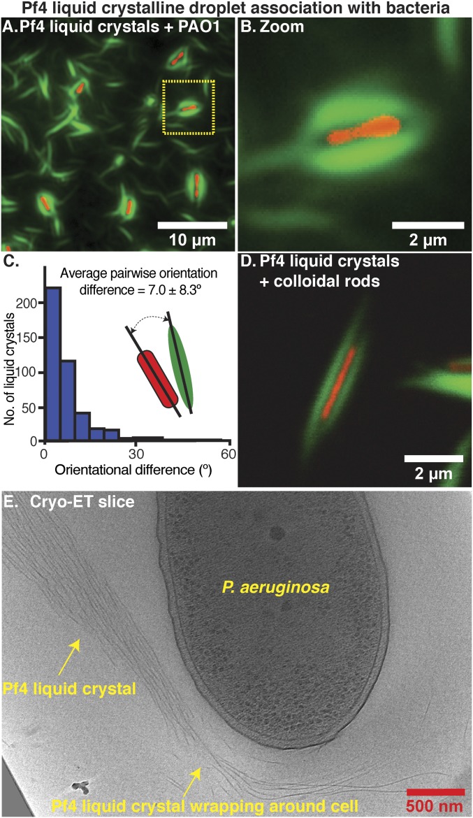
Fig. 5.
Pf4 liquid crystalline droplets form protective sheaths around P. aeruginosa cells. (A) Optical microscopy of the condition from the antibiotic protection assay presented in Fig. 4, containing P. aeruginosa cells, alginate, and Pf4 (Fig. 4A, bar 4, no antibiotic). Transmitted light channel shows bacteria (red pseudocolor) and green fluorescent channel shows Pf4 (green). (B) Zoom of cell shows close association of liquid crystalline droplet around the cell. (C) Histogram of pairwise orientational differences between bacterial cells and associated liquid crystalline droplets from automated segmentation of fluorescence images (n = 417). (D) Colloidal rods (with a similar morphology to bacteria) mixed with Pf4 liquid crystalline droplets show the same encapsulation effect. (E) Cryo-ET slice showing a Pf4 liquid crystal phase associated with and wrapping around a bacterial cell (Movie S5).

