Fig. 6.
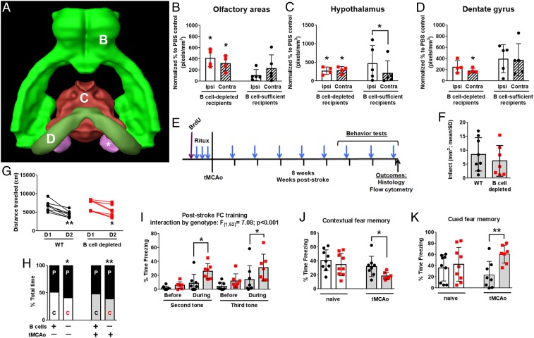
B cell depletion increases general anxiety and spatial memory deficits independent of infarct volume. (A) Brain Explorer surface rendering shows brain regions with B cell diapedesis related to cognitive function, including (B) olfactory areas, (C) hypothalamus, and (D) dentate gyrus, as indicated by letter in the figure. Both B cell-depleted mice (red squares) and mice with endogenous B cells at time of stroke (black circles) show B cell diapedesis in ipsilesional (ipsi, white bars) and contralesional (contra, hatched bars) brain regions. Fold change (y-axes) and significance vs. PBS controls shown unless indicated by brackets. (E) Experimental timeline for behavior testing in uninjured mice (white bars) or after transient middle cerebral artery occlusion (tMCAo; gray bars). (F) B cell depletion did not affect infarct volumes or (G) total movement in the open field. (H) Both uninjured and poststroke B cell-depleted mice spent more time in the periphery (“P”) of the open field compared to the center (“C”) of the field. (I) On the first day of cued and contextual fear-conditioning, poststroke B cell-depleted mice exhibited prolonged freezing during the training tones/shocks. (J) The next day, these same mice exhibited a worse spatial memory. (K) On the last day, B cell-depleted mice again exhibited more freezing during cued memory. Data graphed as mean ± SD. Significance determined by paired t test, Student’s t test, or repeated-measures ANOVA (*P < 0.05, **P < 0.01 vs. nondepleted control or D1 unless indicated by brackets).

