
Figure 3: Ribosome and fMet-tRNAfMet dynamics during maturation of the 70S IC into a 70S EC.
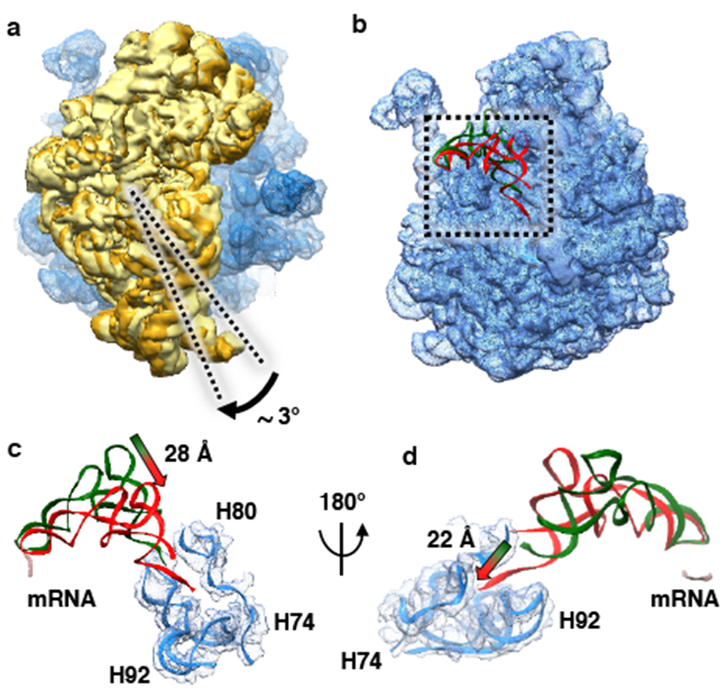
(a) Superposition of the 50S subunits from the 70S IC and 70S EC and comparative analysis of the conformations of the 30S subunit from the 70S IC (pale yellow) and the 30S subunit from the 70S EC (golden yellow). The analysis reveals that the ribosomal subunits from the 70S IC that is initially formed upon 50S subunit joining to the 30S IC transiently acquire a semi-rotated inter-subunit orientation and subsequently undergo an ~3°clockwise rotation, when viewed from the solution side of the 30S subunit, into the non-rotated inter-subunit conformation upon maturation of the 70S IC into a 70S EC. (b) Superposition of the 50S subunits from the 70S IC and 70S EC and comparative analysis of the conformations of fMet-tRNAfMet in the 70S P/I configuration from the 70S IC (green) and fMet-tRNAfMet in the P/P configuration from the 70S EC (red). The start codon of the mRNA is shown in pale pink. The analysis reveals the conformational rearrangements of fMet-tRNAfMet that take place as the 70S IC matures into a 70S EC. (c) A magnified view of the superposition shown in panel (b) reveals that the central domain of fMet-tRNAfMet moves by ~28 Å towards the P site. (d) A 180° rotation of the superposition shown in panel (c) highlights the untangling of the 3’ CCA fMet tail of the fMet-tRNAfMet and its ~22 Å movement into the PTC.