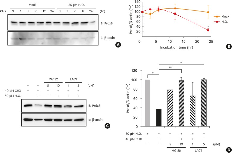
Fig. 4. Degradation of Prdx6 modified by oxidative stress through proteasome in BEAS-2B cells. Degradation of Prdx6 through proteasome was examined by Western blot analysis. (A) BEAS-2B cells were treated with 50 μM H2O2 for 1 hour in HBSS. The cells were washed and incubated in complete medium containing 40 μM CHX for the indicated time. Cell lysates were separated on SDS-PAGE and detected with immunoblotting using anti-Prdx6 antibody. β-actin served as the loading control. (B) The quantified data from (A) are presented as a graph. The ratio of Prdx6/β-actin in mock control was set as 100%. Values are the mean ± SD (n = 3). (C) After 50 μM H2O2 treatment, BEAS-2B cells were incubated in 40 μM CHX with or without MG132 or LACT for 24 hours. (D) The quantified data from (C) are presented as a graph. The ratio of Prdx6/β-actin in BEAS-2B cells treated with H2O2 was set as 100%. Values are the mean ± SD (n = 3).
Prdx, peroxiredoxin; H2O2, hydrogen peroxide; SD, standard deviation; CHX, cycloheximide; HBSS, Hanks' balanced salt solution; SDS-PAGE, sodium dodecyl sulfate-polyacrylamide gel electrophoresis; IB, immunoblotting; LACT, lactacystin.
*P < 0.05 vs. mock treatment for 24 hours; ††P < 0.01 vs. BEAS-2B cells treated with hydrogen peroxide; §§P < 0.01; §§§P < 0.001 vs. BEAS-2B cells treated with hydrogen peroxide and CHX.

