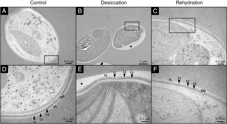
Fig. 5.
TEM micrographs of Csol sections with CW details during a D/R cycle. Rectangles in panels (A–C) indicate the areas shown at higher magnification in panels (D–F), respectively. The asterisk in (B) and (E) indicates a gap between the Epon 812 resin and the CW, probably due to reduced adherence of the collapsed regions to the resin. L1, L2 and L3 indicate the typical three-laminar organization of the CW. FL, fibrillar layer; PM, plasma membrane.

