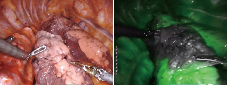
Figure 5.
Using indocyanine green (ICG) pulmonary angiography during anatomical segmentectomy. (A) Normal view of right lower lobe after division of segmental pulmonary arterial branches to superior segment (S6); (B) view with Firefly mode after IV injection of 3 cc ICG clearly demarcates the margin of the non-perfused superior segment (S6).

