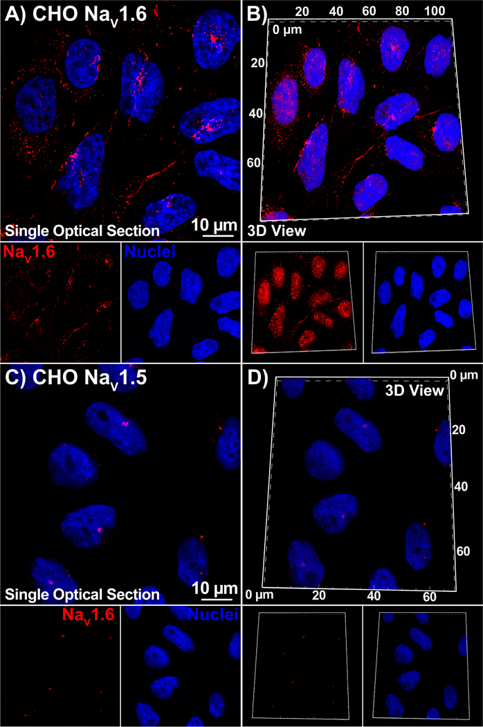
Figure 3. Antibody Specificity in heterologous expression system:

Single optical sections and 3D views of confocal images from CHO cells stably expressing NaV1.6 (A, B) or NaV1.5 (C, D) labeled with our NaV1.6 antibody (raw serum; red) as well as a DAPI nuclear stain (blue).
