
Figure 4. Rabbit Polyclonal anti-NaV1.6 Antibody – Affinity Purified:
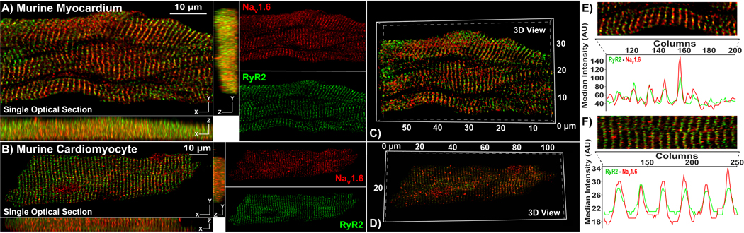
Representative Confocal images from A) myocardial sections, and B) isolated cardiac myocytes immunolabeled using our rabbit NaV1.6 antibody (raw serum; red) and a mouse monoclonal RyR2 antibody (green). Single optical sections (XY) are presented along with orthogonal projections (YZ, XZ) as well as images of individual fluorescence channels. C, D) 3D views of the same samples. E, F) Intensity profiles demonstrate striated pattern of NaV1.6 immunosignals closely associating with RyR2 immunosignals.