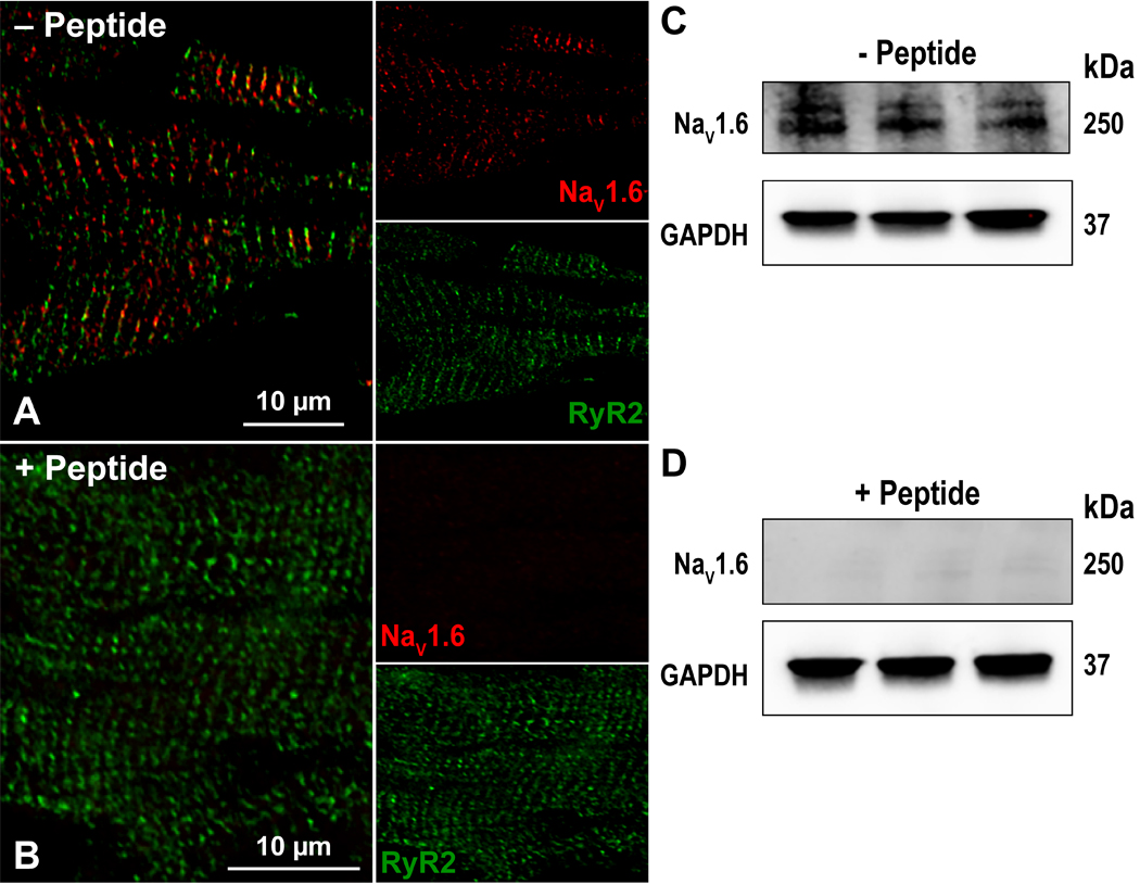
Figure 5. Antibody Specificity – Peptide inhibition.

Confocal images of myocardial sections immunolabeled with for RyR2 (green) and NaV1.6 (red) in the A) absence and B) presence of a peptide corresponding to the NaV1.6 C-terminal epitope. Western immunoblots of whole cell lysates of three WT murine hearts (1 per lane) in the C) absence and D) presence of a peptide corresponding to the NaV1.6 C-terminal epitope. In both cases, GAPDH was detected as a loading control.
