
Figure 6. Antibody Specificity – KO models:
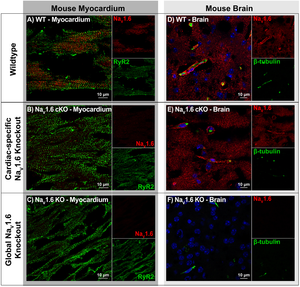
A-C) Confocal images of cardiac sections from WT, cardiac-specific NaV1.6 KO (NaV1.6 cKO) and global NaV1.6 KO mice, labeled for NaV1.6 (red) and RyR2 (green). Clear NaV1.6 immunosignal was observed in WT but not NaV1.6 cKO or NaV1.6 KO. D-F) Confocal images of brain sections labeled for NaV1.6 (red) and β-tubulin (green) show clear NaV1.6 immunosignal in WT and NaV1.6 cKO but not NaV1.6 KO samples.