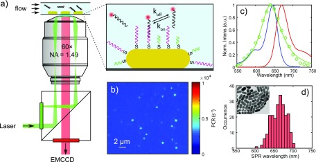
Figure 3.
Single-molecule fluorescence microscopy and single-particle spectroscopy. (a) Schematic illustration of the setup for objective-type total internal reflection fluorescence (o-TIRF) microscopy, with the inset showing the surface functionalization and DNA binding on a single gold nanorod. (b) EMCCD image of immobilized DNA-funtionalized AuNRs immersed in imager solution, with diffraction-limited spots from the one-photon photoluminescence of single AuNRs. (c) Hyperspectral scattering spectrum of a typical single AuNR with a longitudinal SPR wavelength of 640 nm (green) and the ensemble absorption/emission spectra (blue and red) of ATTO 647N in aqueous buffer. (d) The distribution of longitudinal SPR wavelengths measured by single-particle hyperspectral spectroscopy. The inset is a TEM image showinga dried droplet of suspension from which we obtained the size and geometry of the AuNRs.

