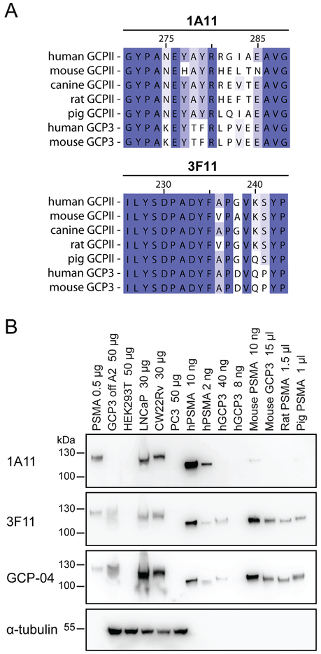
Fig. 1.
Epitope mapping and mAb Western blotting. (Panel A): Alignment of the epitopes on PSMA from different species recognized by the mAbs 1A11 and 3F11 as revealed by peptide scanning. Panel B: Purified ectodomains of human PSMA and several orthologs/paralogs, cell culture supernatants as well as cell lysates were separated by reducing 10% SDS–PAGE, electrotransferred onto a PVDF membrane, and probed with individual mAbs. Lanes: 1. human PSMA-overexpressing HEK293T/17 lysate (0.5 μg); 2. GCP3 overexpressing HEK293T/17 lysate (50 μg); 3. HEK293T/17 lysate (50 μg); 4. LNCaP lysate (30 μg); 5. CW22Rv1 lysate (30 μg); 6. PC-3 lysate (50 μg); 7. human PSMA (10 ng); 8. human PSMA (2 ng); 9. human GCP3 (40 ng); 10. human GCP3 (8 ng); 11. mouse PSMA (10 ng); 12. mouse GCP3 (cell culture supernatant; 15 μl); 13. Rat PSMA (cell culture supernatant; 1.5 μl); 14. pig PSMA (cell culture supernatant; 1 μl).

