Fig. 2.
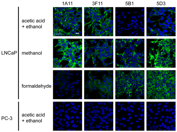
PSMA detection on LNCaP (PSMA-positive) and PC-3 (PSMA-negative) cell lines by immunofluorescence microscopy. Individual cell lines were fixed on glass coverslips using three different fixation protocols and probed with tested mAbs (20 μg/ml), followed by detection with a secondary antibody conjugated with Alexa Fluor 488 (green channel). Under these varying conditions distinct intensities of both cytoplasmic and plasma membrane staining were observed on LNCaP cells. The PSMA-negative PC-3 prostate cell line revealed no staining (cells fixed by acetic acid + ethanol are shown as an example). Nuclei were visualized with DAPI (blue channel); scale bar: 20 μm.

