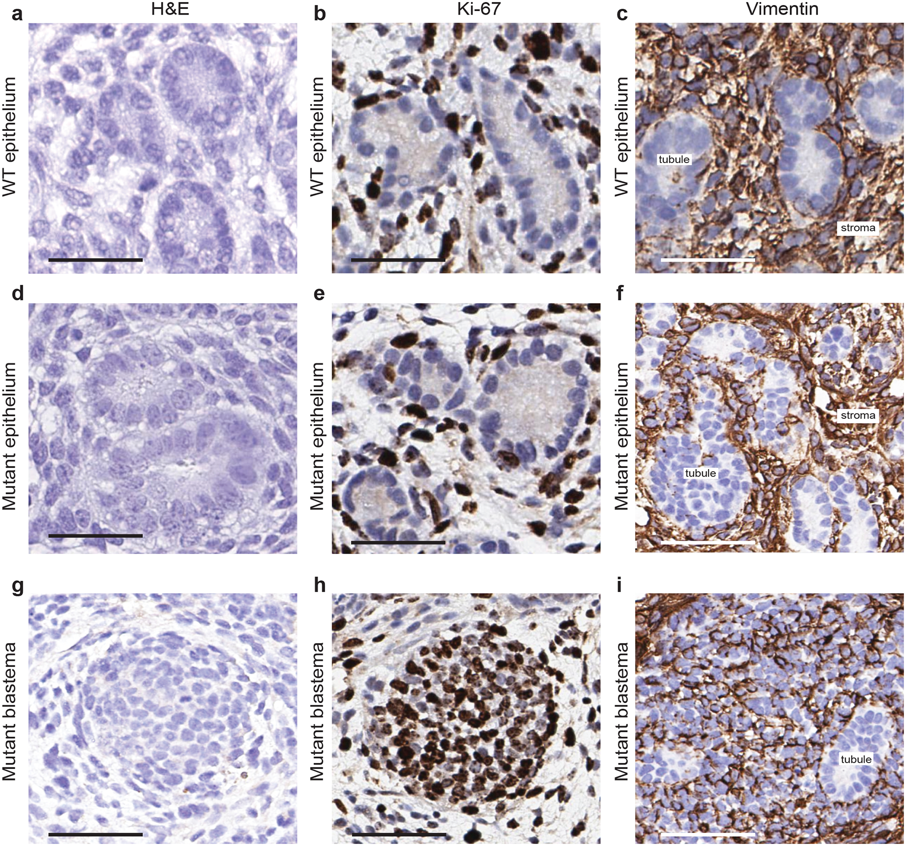
Extended Data Fig. 3 |. Characterization of ENL-mutant kidney structures.

a, d, g, Representative haematoxylin and eosin staining of the indicated kidney structures. b, e, h, Representative immunohistochemistry staining of the indicated kidney structures for the proliferation marker Ki-67. c, f, i, Representative immunohistochemistry staining of the indicated kidney structures for the mesenchymal marker vimentin. In panels c, f, the vimentin-positive cells shown are stroma cells. In panel i, the vimentin-positive cells shown are mostly blastema components. a–c, WT epithelium; d–f, mutant epithelium; g–i, mutant blastema. All experiments were repeated twice with similar results. Scale bars, 50 μm.
